Janus Archives
.Janus the Greek god of duality, marking the beginning and the end, or the past and the future, is an apt name for the Archives at Seth GS Medical College, which has a collection of historical manuscripts, books, photographs, tools, equipment and furniture that have been preserved for future reference.
Archives are essential for understanding the past and present. They provide valuable information about historical events, developments, and can also aid research. They are an important resource for scholars, researchers and interested individuals.
Archives are also essential for preserving institutional heritage and maintenance of the Gosumec identity. Their preservation and management are critical for ensuring that they remain accessible for generations to come.
Dr. Pragna Pai’s support and funds enabled Dr. Manu Kothari, Dr. Sunil Pandya, Artist Avinash Marathe and Dr. Lopa Mehta to set up the Janus Archives on Foundation Day in 1991. The archives was relocated during the renovation of the college building, and has been in its current location since 2013.
Dr. Lopa Mehta, the curator of the Archives, is ably supported by Dr. Swarupa Bhagwat and the student secretaries. Together, they are dedicated to preserving the history of the institution.
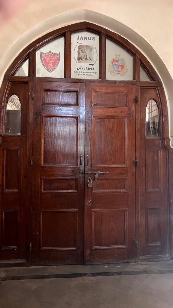
Janus Archives, located on the ground floor of the medical school building, adjacent to the Pathology museum, contains historical relics enabling a glimpse into the history of medicine, knowledge, culture, and society in India. Some of these antiquarian books are over 200 years old, neatly stored in cabinets and have withstood the test of time.
Evolution of EKG machines
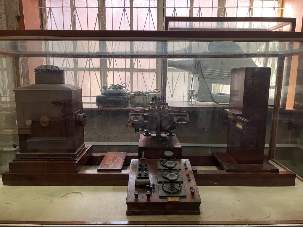
First ever EKG machine imported in India (late 1940s) which was also used to record the EKG of Mahatma Gandhi.
The galvanometer made electrocardiography practical, creating a new branch of medicine and even a new industry. Einthoven (1913) and Lewis (1920) dominated the early years of electrocardiography. The period following 1920 was influenced largely by Wilson. None did as much to advance EKG knowledge as did Wilson. The interest shifted to the theory of the EKG, abnormalities of wave form and of EKG leads.
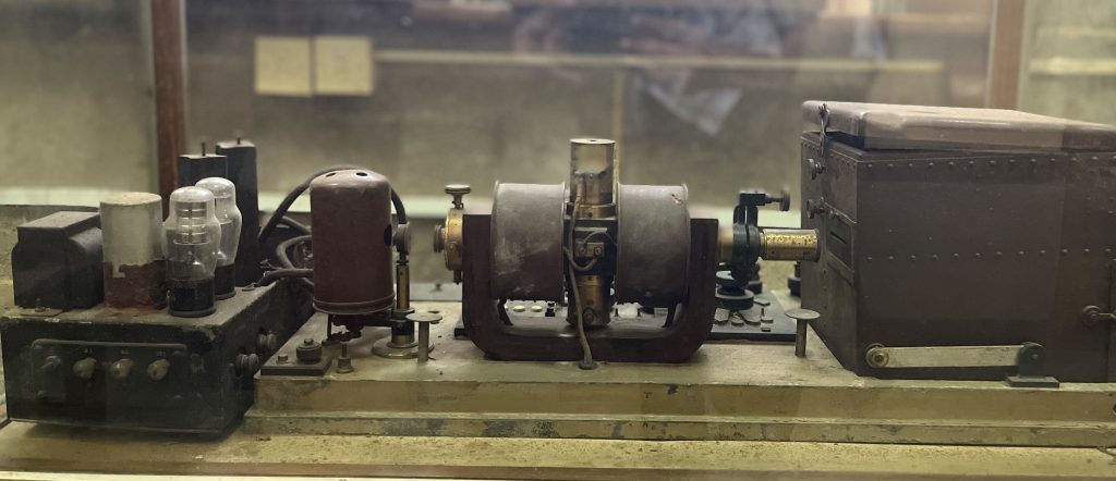
Boulitte Electrocardiograph, manufactured in France, 1900-1030. EKGs are graphical records of the electrical activity of the heart produced via an instrument called a galvanometer. Here the galvanometer is seen in the center. Electrodes placed upon the body pick up on minute electrical impulses. These cause a string within the galvanometer to move. Movement is recorded photographically to make a tracing. A plate camera can be seen in the rectangular box at the end. This EKG machine was made by a French company, Boulitte.
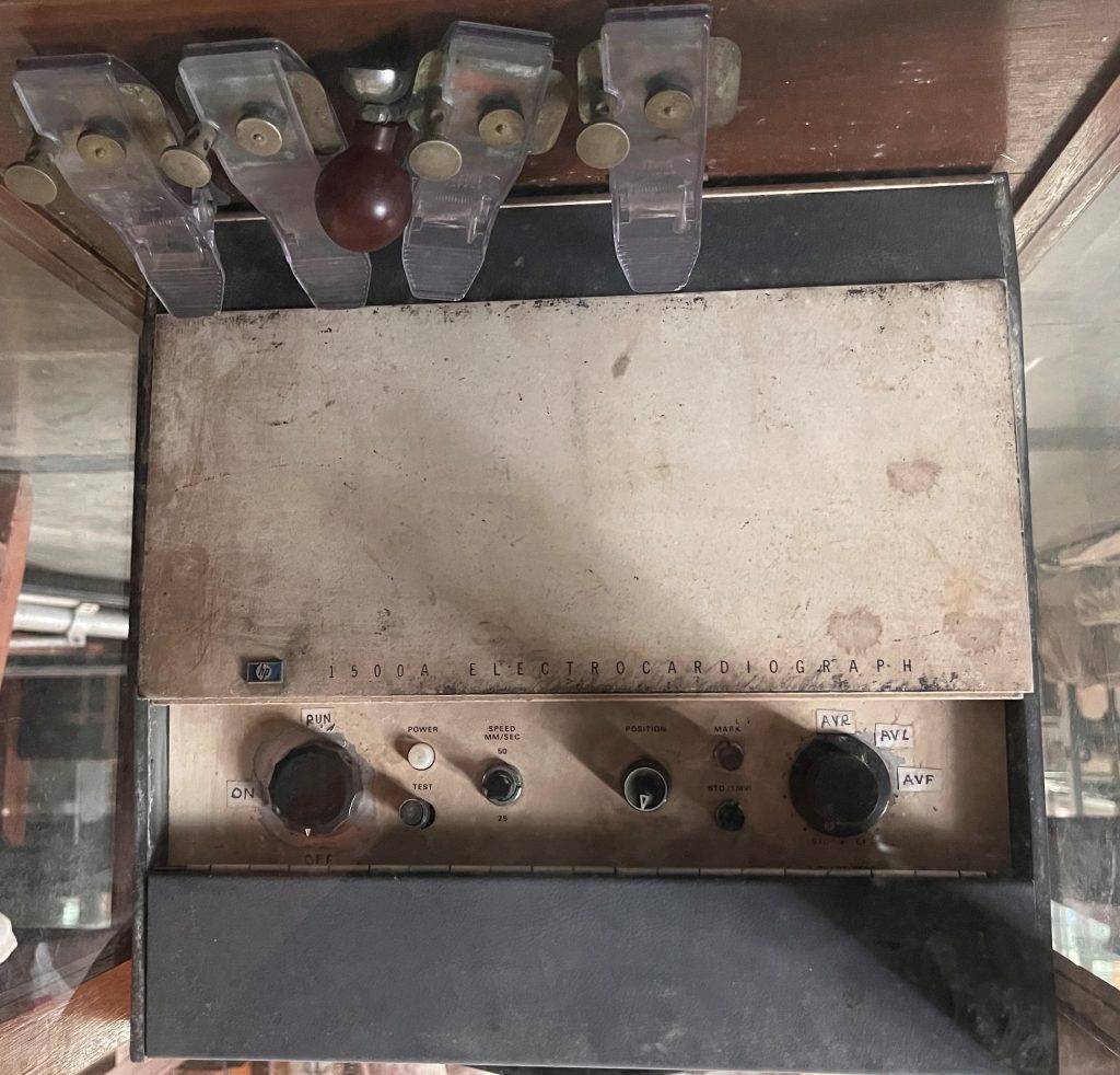
1961, The Sanborn Company, early EKG Machine makers was acquired by Hewlett Packard who continued the manufacture of EKG machines.
Shown is the HP 1500A machine introduced into KEM Hospital in 1960-70s.
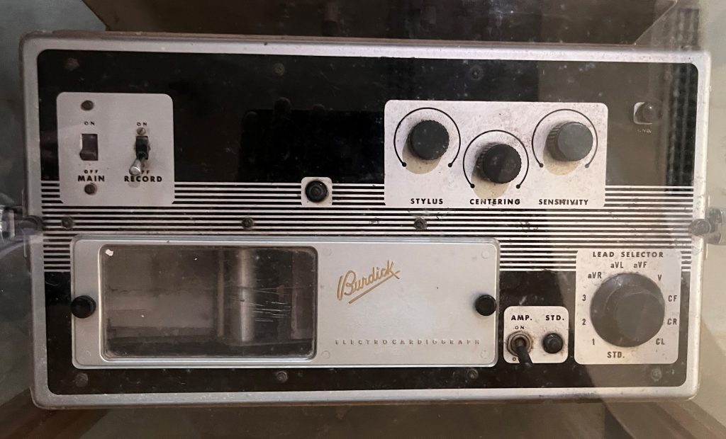
THE EK-2 Portable EKG machine manufactured by Burdick Corporation (1970s and 1980s) enabled recordings in a portable format, allowing medical professionals to take EKG readings in a variety of settings.
Stereotaxy Apparatus
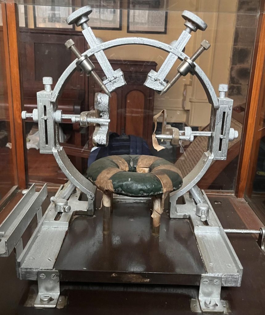
This stereotaxy apparatus used by Dr. Homi Dastur and Dr. Anil Desai at KEM, was fabricated based on the Japanese original, soon after Dr. Desai visited Prof. Narabayashi in 1962.
Though originally trained as a neuropsychiatrist, Prof. Narabayashi is best known for his pioneering work in the management of movement disorders, especially neurosurgical treatment of Parkinson’s disease, till levodopa discovery in 1968 changed the treatment paradigm. His stereotactic apparatus was assembled based on drawings of the equipment used in the US by Dr. Victor Horsley and Dr. Robert H. Clarke.
The origin of stereotactic neurosurgery for movement disorders was preceded by the infamous leucotomy, subsequently called lobotomy, a psychosurgery, first performed in 1935 by Portuguese Neurologist Prof. Antonio Egas Moniz, for which he received the Nobel prize in 1949!
Portable ICU monitor
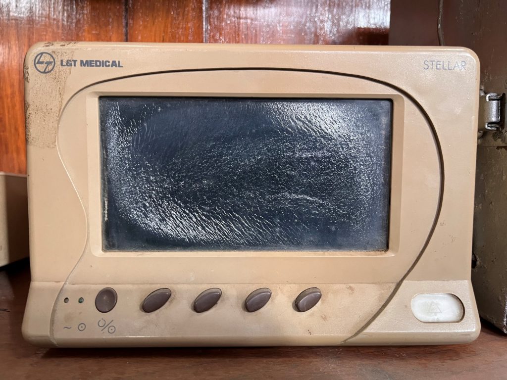
The first portable Patient monitoring system in the Cardiothoracic ICU (Dr. P.K.Sen era) led to enhanced patient safety in the perioperative unit. This “modern” ICU monitor replaced large and bulky vintage models.
The vintage models with Cathode-Ray Tube (CRT) displays, showed waveforms and numerical values for each vital sign. These stand-alone units required a dedicated space in the ICU with a trained technician to operate them.
Pulse Oximeter
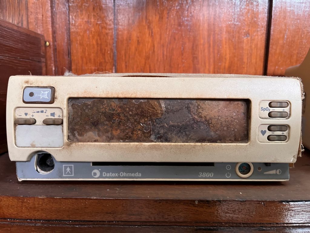
Takuo Aoyagi, a Japanese inventor, an electrical engineer, developed the pulse oximeter in the early 1970s (centuries after the use of anesthesia for medical procedures).
Awareness of the importance of anesthesia safety in the US, due to academic foresight and media attention, in combination with excellence in technological innovation, led to widespread use of pulse oximetry around the world. Now, miniaturized pulse oximeters have found their way in almost every home in the US (Covid era).
Even today, we are still searching for the theory of pulse oximetry.
Oxygen tank
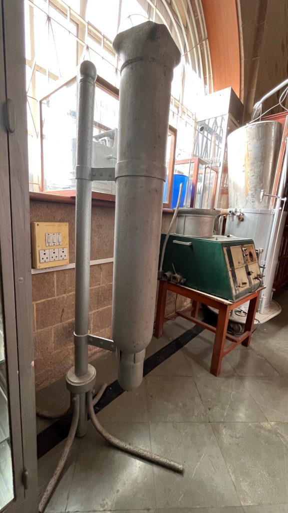
A “portable” oxygen storage vessel held under pressure in gas cylinders. These cylinders have been miniaturized for current day use, including portable tanks and concentrators for individual use.
Kala Ghoda
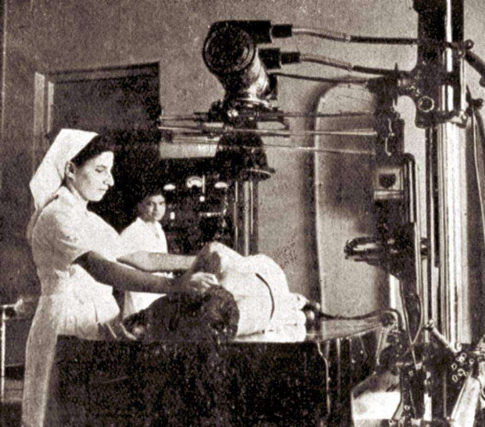
The GE X-ray machine nicknamed “Kala Ghoda” was a steady presence in the Radiology Department’s OPD no. 10 from its installation in 1944-45 until its retirement in the early 1970s.
The machine’s black color and solidity inspired its nickname, which means “black horse” in Hindi.
The Engineering Department was responsible for maintaining the machine, which did not have a light beam diaphragm but did have an attachable small cone for various cone-down exposures.
Iron Lung
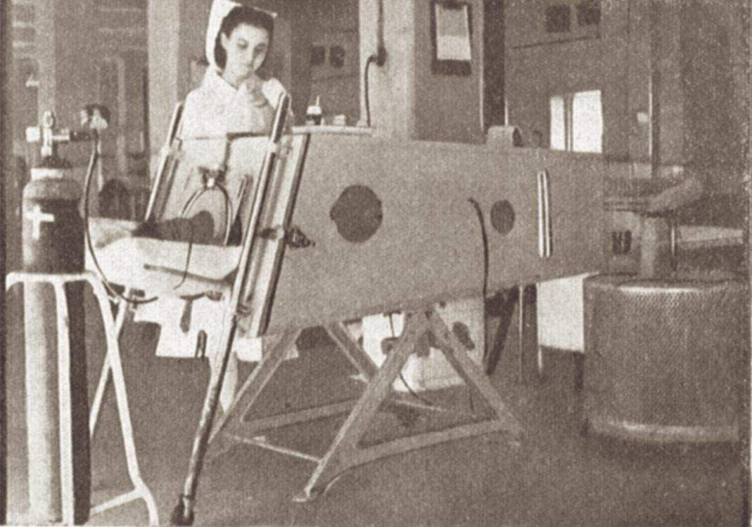
The iron lung is a negative-pressure ventilator that was widely used during the polio epidemics of the 1940s and 1950s.
Patients who contracted polio and lost the ability to breathe on their own would be placed in an iron lung, which would help them breathe by creating a negative pressure around their chest. Patients would typically spend two weeks in an iron lung while they recovered, but some would need to remain in the machine for much longer.
The development of the polio vaccine in the 1950s led to a dramatic decline in the use of iron lungs, as the vaccine effectively prevented polio from spreading.
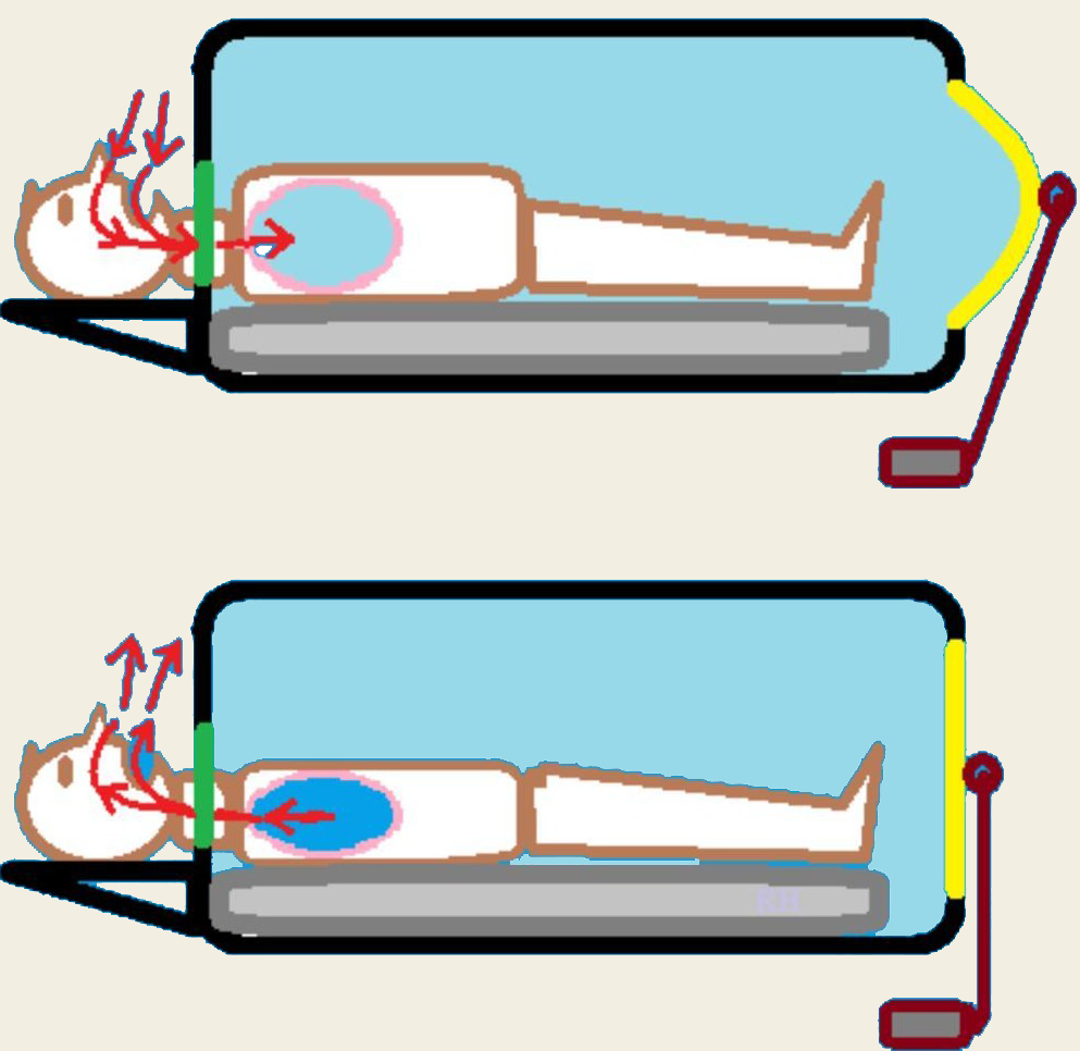
Philip Drinker and Louis Agassiz Shaw invented the first iron lung at Harvard School of Public Health. Later in Australia, the Both respirator which was made of plywood and cheap, easy to construct, transport and was in use within one hour of production!
The rhythmic “whoosh” of air from the iron lung, created by the bellows as they moved air in and out, became a reassuring sound of patients breathing.
One of the biggest problems for patients was boredom. A mirror could be attached above the patient’s head, so they could see what was happening around them. They could also read books suspended in front of their faces if someone turned the pages for them.
Tetanus
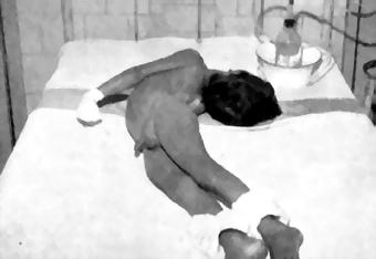
Opisthotonus
Tetanus’ Arched Back Phenomenon
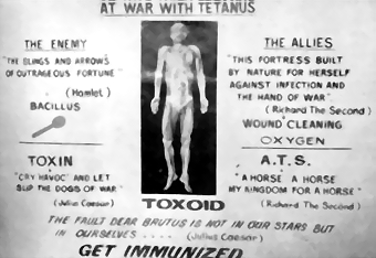
Allies-Anti toxin serum combat vs. Tetanus Neurotoxin
This ailment features initial “lockjaw” leading to stiffness, painful contractions, and the arched opisthotonus posture. Even minor triggers induce spasms impacting breathing, often causing respiratory failure—a common cause of death, sometimes worsened by secondary infections.
Key treatment is antitoxin serum (ATS), containing antibodies counteracting tetanus toxins. Comprehensive management includes human tetanus immunoglobulin, wound care, muscle relaxants, and intensive support like ventilation. The fatality rate dropped impressively from 100% to below 20%.
Vintage Microscope
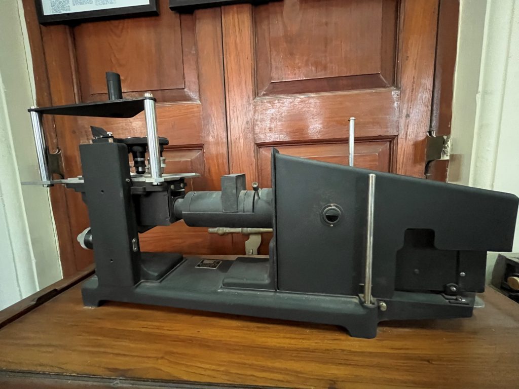
Progressing from the Nimrud lens (710 BC), in 1676, Antonie van Leeuwenhoek built a simple microscope with one lens to examine cells and bacteria that allowed for magnifications of up to 270 times.
Continued evolution of the microscope to the current era of super microscopes (super-resolved fluorescence microscopy) which now allow to “see” matter smaller than 0.2 micrometers.
Vintage PFT
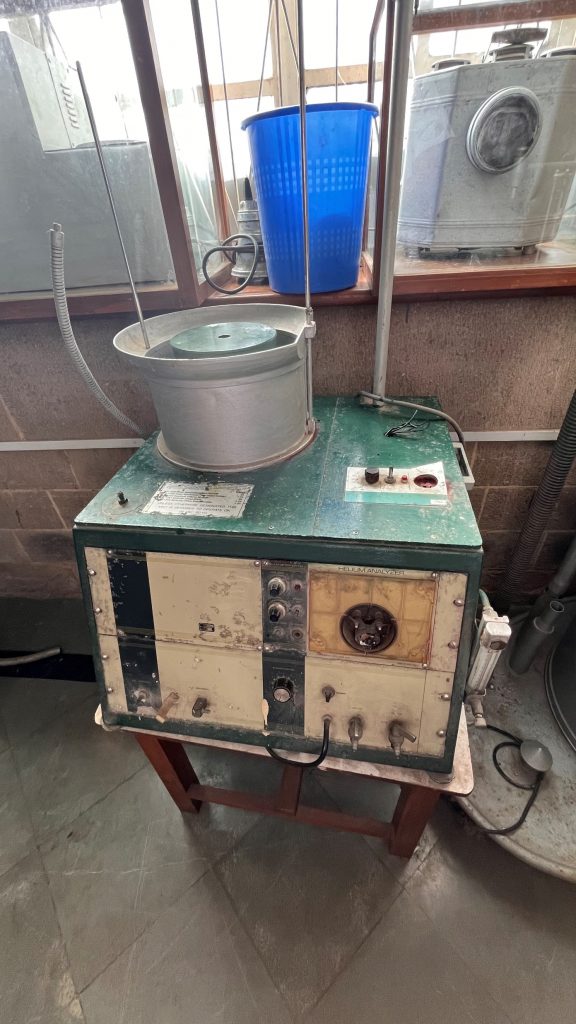
Called the capacity of life, John Hutchison (1839), an engineer-physician, hired by the Brittanic Assurance Society, a new firm into the nascent London life insurance market was the creator of early Pulmonary Function Tests. This was an advancement from the actuarial tables used by life insurance companies to predict insurance risk. He spent the remainder of the 1840s continuing to collect vital capacity data, publish his findings, and enhance the body of knowledge of pulmonology.
By 1852, bored with life in London, he packed up his belongings, and moved to literally the other side of the world, settling in Australia (Gold Rush), leaving his academic interest back in his London office.
He moved once again to the island of Fiji (1861), where he unfortunately contracted dysentery after arriving and died the same year, leaving behind a vital legacy that continues to influence devices to this day. This vintage pulmonary function device is in the natural evolution to the current miniatures used in offices around the world.
Epidiascope
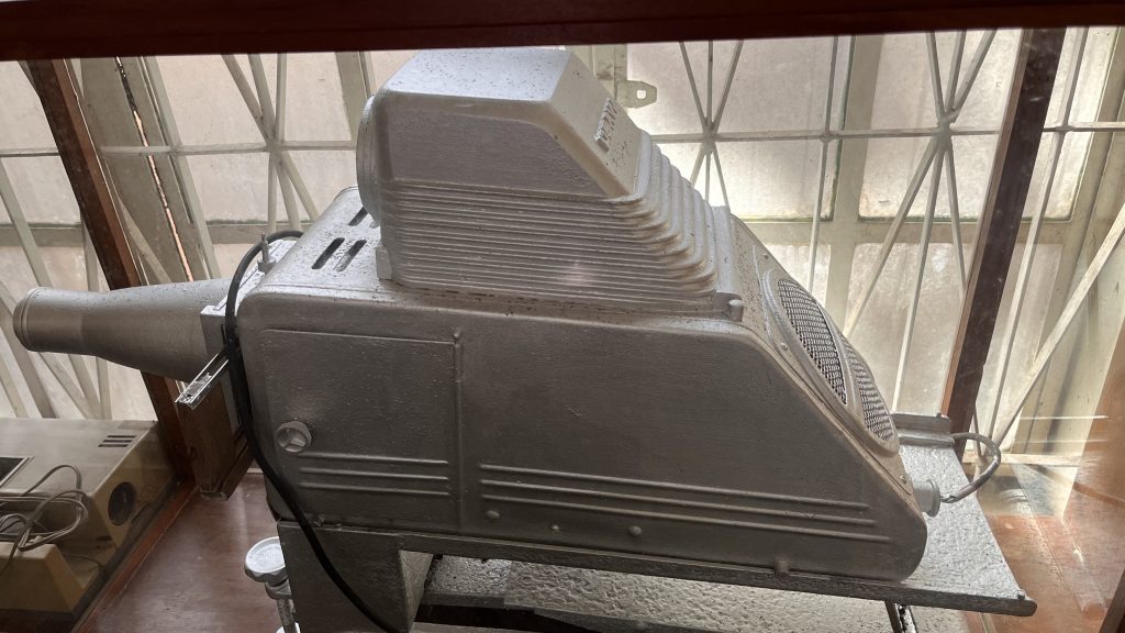
Epidiascope, a teaching aid, is an optical projector capable of producing images of both opaque and transparent objects. It consists of an episcope and a diascope. In the epi-position, it can project opaque and flat objects such as textbooks, journal pages and drawing.
Vintage Projector
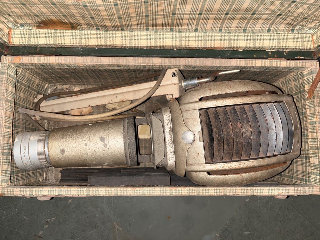
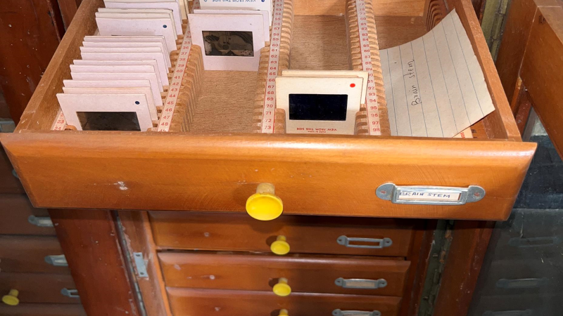
Progressing from Magic Lanterns was the slide era. Introduction of color slides was the last major advance in slide technology.
Moving from the analog era, we have progressed to digital images using overhead projector-screens and digital technology as teaching aids.
Vintage Typewriter
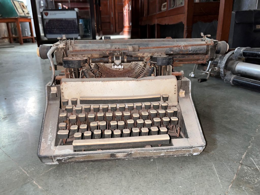
Type-Bar Typewriters were the most common kind of typewriter. A mechanical device to produce printed characters on a piece of paper by typing individual keys. The paper was rolled around a cylinder mounted on a carriage.
Used at KEM to record manuscripts, thesis and official documents, these are antique relics of the 19th and 20th century, which have been replaced by modeled current keyboards which use the same key format, including the shift key. This device has been replaced with a computer-keyboard-printer combination.
Contributed by Prajwal Varak
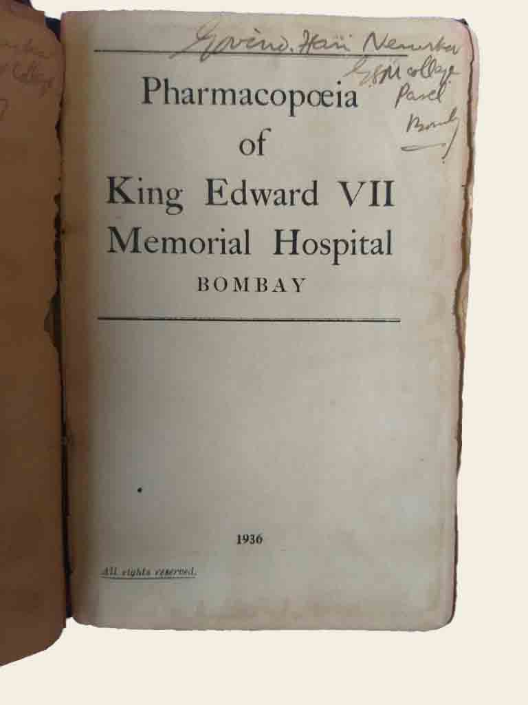
Pharmacopeia, 1936
By clicking the image, Delve into the extraordinary and rare original pharmacopeia of KEM Hospital Bombay, meticulously preserved since 1936.
This fascinating historical treasure offers an unparalleled opportunity to unravel the medical practices and pharmaceutical knowledge of that bygone era, providing invaluable insights into the evolution of healthcare.
Contributed by Dr. Malini Nadkarni
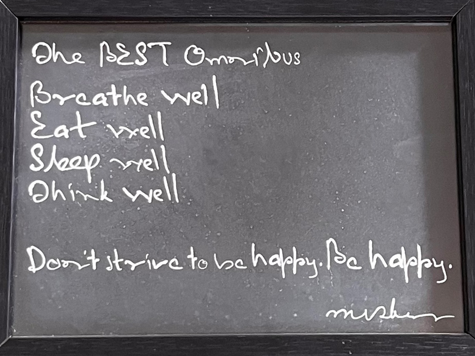
Be happy
